Background
Concussion has received increasing focus in recent years, especially with respect to athletes and contact sports participation. Study of the impact on certain professional athletes’ brains is one area of focus, given their long-term exposure to repetitive impacts with an inherent risk of concussion; another is the impact on the developing brain of children and adolescents who participate in risky contact sports while their brains are still maturing. The maturing brain may be uniquely susceptible to long-term change from concussion.
The bulk of the deep parts of the brain are formed of white matter, the tissue that allows messages to pass between the areas of neurons known as grey matter. By using high field-strength MRI and sophisticated analytical methods, it is possible to detect prolonged abnormalities in the white matter of the brain that would otherwise be invisible using a normal clinical MRI scan. Are there still changes occurring in the adolescent brain even after the clinical evaluation and standard assessment has otherwise approved athletes to return to sport?
The Research
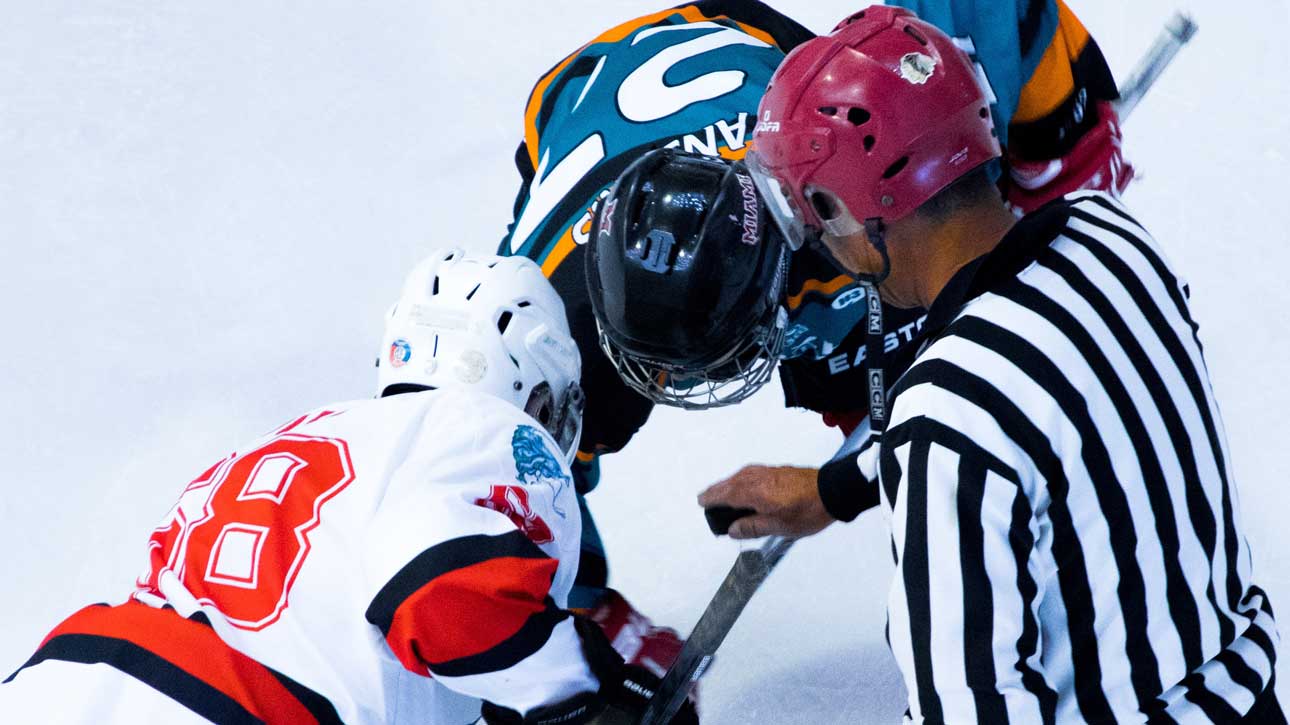
We recruited 17 concussed male hockey players from Bantam leagues (aged 11-14, when body checking is first introduced) and a control group of 26 age-matched players. We evaluated the concussed players over time (24-72 hrs after an injury and again at 3 months when they were approved for return to play) using a variety of advanced MRI techniques and compared that data to the control group.
The Findings
We detected abnormalities at both sets of scans in the concussed players, suggesting those abnormalities can be quite prolonged, past the point at which the standard clinical tests have approved the player to return to play. The abnormalities were diffusion-related in the white matter of the brain, changes in connectivity and decreases in the metabolites (small molecules required for normal growth and development through metabolic processes inside the brain) in the prefrontal white matter.
Hyperconnectivity was detectable at three months after injury compared to both the control group and the 24-72 hr scans – hyperconnectivity refers to more highly correlated brain activity between areas of the cortex and has been proposed as a characteristic of recovery and compensation for disruption within the white matter of the brain.
Next Steps
Our results in this study suggest that this adolescent population may require longer recovery periods after a concussion. It has shown that changes persisted well after a player’s clinical scores had returned to normal and they had been cleared to play. Further research is required to understand the longer-term impact of early brain injuries but this work will help to develop a better clinically-relevant, objective measure for concussion diagnosis than current standard tests. We are currently analyzing longitudinal data from the Western women’s varsity rugby team, as far as six months after a concussion, to determine if and for how long these brain changes persist.
Anyone involved in contact sport in the adolescent age range should be aware of the increased recovery time this work suggests.
Key Points
Changes continue to occur in a concussed brain even after standard clinical tests have returned to normal. Damage in the very long fibre tracks in the brain of concussed players can be detected up to three months after the concussion and after the individuals have been approved for return to athletics. It is also possible to detect ‘hyper-connectivity’ in the brain, suggesting the brain is still trying to compensate for the concussion.
Publication
Neurology.org October 2017 http://n.neurology.org/.../2157
Western Researchers
Kathryn Y. Manning
Amy Schranz
Robert Bartha
Gregory A. Dekaban
Christy Barreira
Arthur Brown
Lisa Fischer
Kevin Asem
Timothy J. Doherty
Douglas D. Fraser
Jeff Holmes
Ravi S. Menon
Author: Western BrainsCAN
Date: 2022/03/08
Source: https://brainscan.uwo.ca/research/research_summaries/KAMA0518-R.html


